
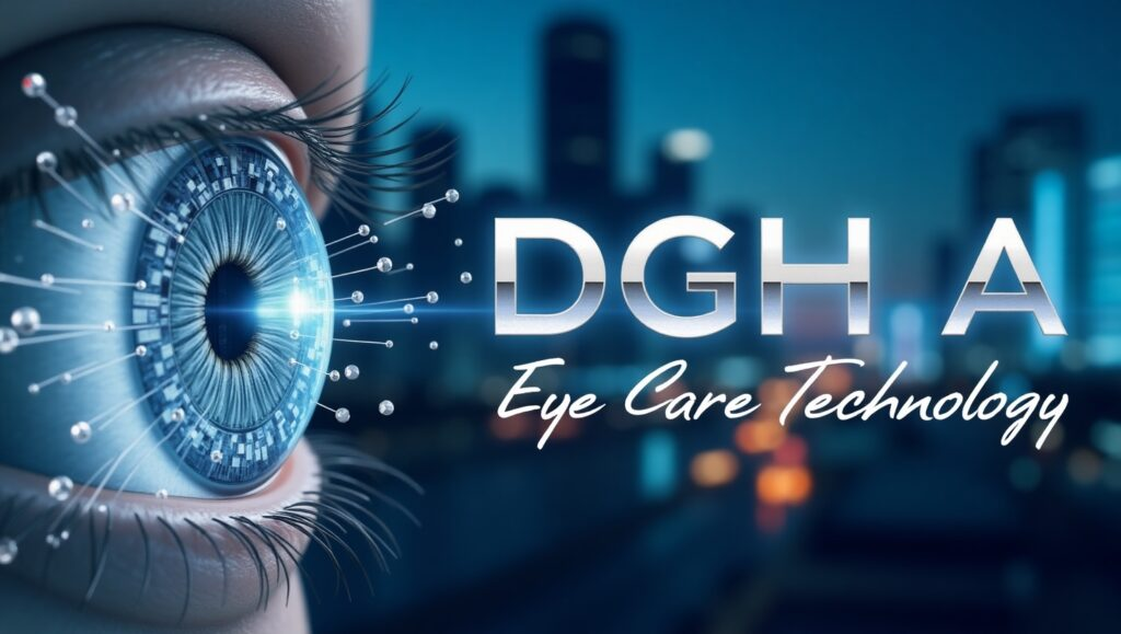
The DGH A, or DGH Scanmate A, is a cutting-edge ultrasound device designed to provide high-resolution eye scans, revolutionizing the field of ophthalmology. This device is not only instrumental in diagnosing eye conditions but also serves as a vital tool for accurate measurements and real-time feedback. By offering non-invasive imaging capabilities, it ensures a quick and effective examination without the need for surgery.
In this article, we will explore the DGH A, its features, applications, and the profound impact it has on the ophthalmic industry. We will also discuss the advancements in ultrasound technology and how this device stands out among its competitors. Whether you’re a healthcare professional, researcher, or someone interested in ophthalmology, this article will provide valuable insights into the DGH A.
Understanding the DGH A: What is it?
The DGH A (often referred to as the DGH Scanmate A) is an advanced ultrasound device primarily used in ophthalmology for eye imaging. It is equipped with high-frequency sound waves that produce detailed images of the structures inside the eye. The device’s primary purpose is to measure various parameters, including the axial length of the eye, which is crucial in diagnosing conditions like myopia, hyperopia, and astigmatism.
Unlike traditional imaging methods, which may require surgery or invasive procedures, the DGH A provides a non-invasive solution for obtaining accurate and high-quality scans. This is particularly valuable for patients who require frequent eye checkups or those with underlying conditions that need constant monitoring.
Also Read: HydraHD: The Future of High-Quality Streaming
Key Features of the DGH A
The DGH A stands out due to its state-of-the-art technology and user-friendly interface. Below are some key features that make this device indispensable in modern ophthalmology:
1. High-Resolution Imaging
The DGH A utilizes advanced ultrasound technology to capture high-resolution images of the eye. This allows doctors to assess the eye’s internal structure with precision, offering an in-depth look at the retina, lens, and other crucial components.
2. Non-Invasive Procedure
One of the major advantages of the DGH A is that it provides non-invasive imaging, eliminating the need for surgical procedures or uncomfortable tests. This makes it a suitable option for routine eye exams and long-term monitoring of chronic eye conditions.
3. Real-Time Feedback
With real-time feedback capabilities, the DGH A enables healthcare professionals to make timely decisions based on up-to-date information. This is crucial for accurately diagnosing eye diseases and conditions such as cataracts, glaucoma, and macular degeneration.
4. Accurate Measurements
The device excels in accurate measurements, particularly for the axial length of the eye. This is a vital metric in understanding the eye’s optical properties and is often used to assess the risk of certain refractive errors and to plan for surgeries such as cataract surgery.
5. Portable and Easy-to-Use
Despite its advanced technology, the DGH A is compact and portable, making it suitable for both clinical and field use. Its user-friendly interface ensures that healthcare providers can operate it with minimal training.
6. Comprehensive Diagnostic Tool
The device is capable of performing a variety of diagnostic tests. It can measure axial length, anterior chamber depth, and lens thickness, all of which are critical in determining eye health and diagnosing refractive errors.
Applications of the DGH A in Ophthalmology
The DGH A has broad applications in ophthalmology, primarily in the diagnosis and management of various eye conditions. Below are some of the most common uses:
1. Refractive Error Diagnosis
By measuring the axial length and other eye parameters, the DGH A helps in diagnosing refractive errors such as nearsightedness (myopia), farsightedness (hyperopia), and astigmatism. It plays a crucial role in pre-surgical assessments for refractive surgeries like LASIK.
2. Cataract Surgery Planning
The DGH A is commonly used in cataract surgery planning to measure the eye’s internal dimensions. These measurements are essential for determining the appropriate intraocular lens (IOL) to be implanted during surgery, helping improve patient outcomes and reducing complications.
3. Glaucoma Management
In glaucoma management, the DGH A can be used to assess the optic nerve head and other key eye structures, helping in the diagnosis of glaucoma and monitoring its progression.
Also Read: Overproduktion: Causes, Impacts, and Solutions
4. Macular Degeneration and Retinal Disorders
The DGH A is valuable for monitoring diseases like macular degeneration and other retinal disorders. By capturing high-resolution scans of the retina, ophthalmologists can track disease progression and determine the effectiveness of treatments.
5. Post-Surgical Monitoring
After eye surgeries like cataract surgery or vitrectomy, the DGH A helps monitor the healing process and ensure the correct positioning of the intraocular lens (IOL). It is also useful for tracking any post-operative complications.
How the DGH A Benefits Patients
The introduction of high-tech devices like the DGH A has significantly improved patient care in ophthalmology. Here are some key benefits for patients:
1. Faster Diagnosis
With real-time imaging and instant feedback, the DGH A allows for faster diagnoses of eye conditions. This leads to quicker intervention, which is especially important in conditions like glaucoma and diabetic retinopathy, where early detection can prevent vision loss.
2. Reduced Need for Surgery
Since the DGH A is a non-invasive imaging tool, it reduces the need for surgery or more invasive procedures. This means fewer risks for patients and a more comfortable experience overall.
3. Long-Term Eye Health Monitoring
The DGH A is invaluable for long-term monitoring of chronic eye conditions. Its ability to perform frequent, accurate scans allows for continuous tracking of a patient’s eye health and timely adjustments in treatment.
4. Enhanced Treatment Outcomes
With accurate and timely information, doctors can make better decisions regarding treatment plans. This leads to improved treatment outcomes and higher satisfaction for patients.
The Future of DGH A and Ultrasound Technology in Ophthalmology
As technology continues to evolve, the DGH A and similar ultrasound devices are likely to become even more advanced. Future developments may include integration with artificial intelligence (AI) to provide even more accurate diagnostics, as well as enhancements to the imaging quality, making it possible to detect even subtle changes in the eye’s structure.
Additionally, the portability of devices like the DGH A means that they could be used in more remote locations, extending access to eye care in underserved regions. This would be particularly beneficial for patients in rural or low-resource settings, where access to specialized care is often limited.
Also Read: Transds: The Next Evolution in Dynamic Digital Systems
FAQs About the DGH A
1. What makes the DGH A different from other eye imaging devices?
The DGH A offers non-invasive, high-resolution imaging with real-time feedback, making it more efficient and less uncomfortable for patients compared to other imaging devices.
2. Can the DGH A be used in routine eye exams?
Yes, the DGH A is perfect for routine eye exams as it provides quick, accurate scans without the need for invasive procedures.
3. Is the DGH A suitable for all types of eye conditions?
While the DGH A is primarily used for refractive errors, cataract surgery planning, and monitoring retinal diseases, it is also effective in managing other chronic eye conditions such as glaucoma and macular degeneration.
4. How does the DGH A improve patient comfort?
The DGH A is a non-invasive device, meaning there is no need for surgery or uncomfortable tests. It provides a painless and quick examination, which is particularly beneficial for elderly or anxious patients.
5. Can the DGH A be used for pediatric patients?
Yes, the DGH A is safe for use with pediatric patients, as it provides a gentle, non-invasive way to assess eye health without causing discomfort.
6. How often should patients use the DGH A for monitoring?
The frequency of use depends on the patient’s specific condition. For chronic conditions like glaucoma or macular degeneration, regular monitoring may be necessary to track disease progression and adjust treatment plans.
7. Does the DGH A require specialized training to operate?
While the DGH A is designed to be user-friendly, healthcare professionals do require basic training to operate the device effectively. However, the device’s intuitive interface makes it easy to use even for those with limited experience.
Conclusion
In conclusion, the DGH A represents a significant advancement in eye care technology. Its ability to provide high-resolution, non-invasive scans with real-time feedback makes it a game-changer in ophthalmology. Whether used for diagnostic purposes, surgical planning, or routine monitoring, this device ensures that healthcare providers can deliver the best possible care to their patients, leading to improved outcomes and better overall eye health.